Hendrik Bary · 1672 · 28 plates
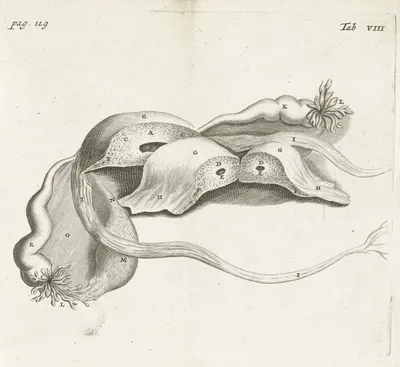
Anatomical detail image of various female genitalia (1672)
1672
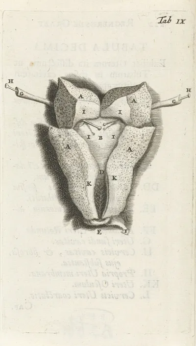
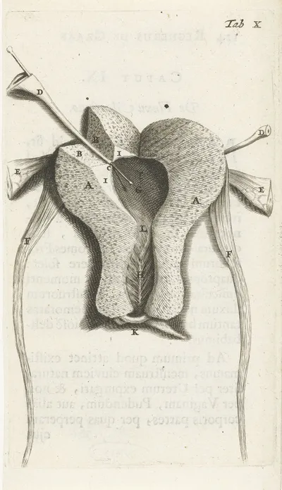
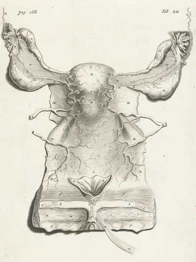
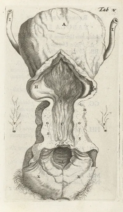
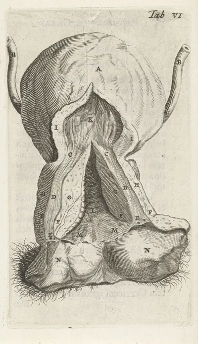
Anatomical drawing of the inside of the uterus. (1672)
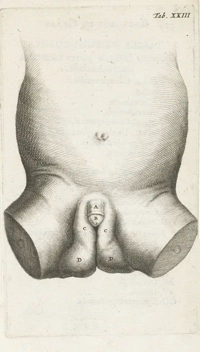
Anatomical illustration of a child’s penis (1672)
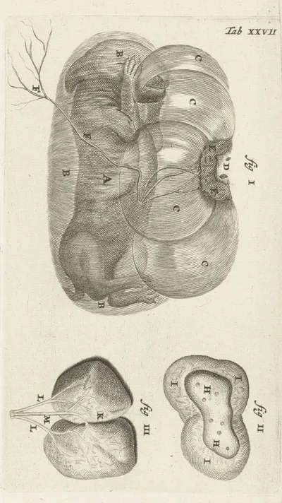
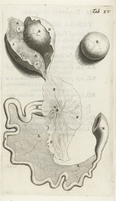
Anatomical illustration of a rabbit fetus in the uterus and the placenta (1672)
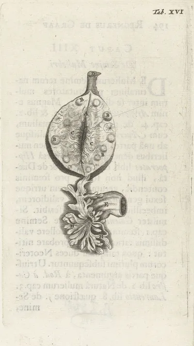
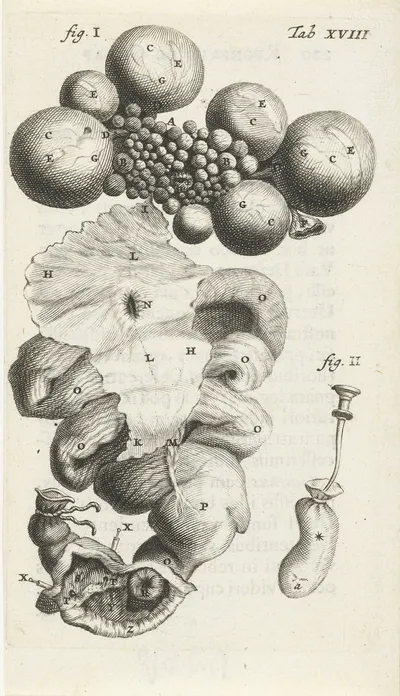
Anatomical illustration of an ovary (1672)
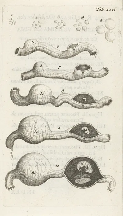
Anatomical illustration of growth stages of the embryo in the uterus (1672)
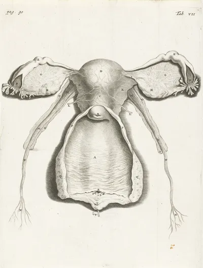
Anatomical illustration of the cervix, uterus, fallopian tubes and ovaries (1672)
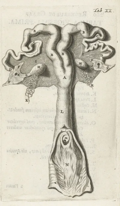
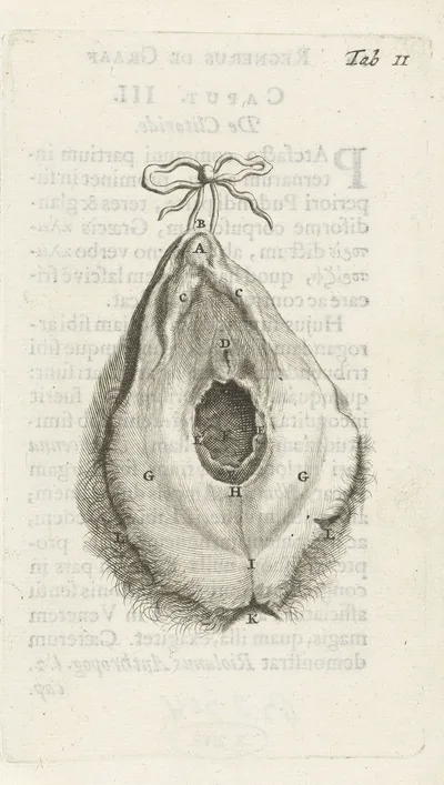
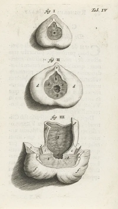
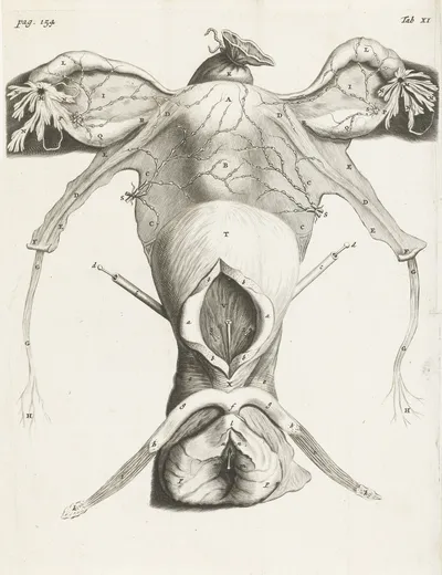
Anatomical illustration of the clitoris, vagina and uterus (1672)
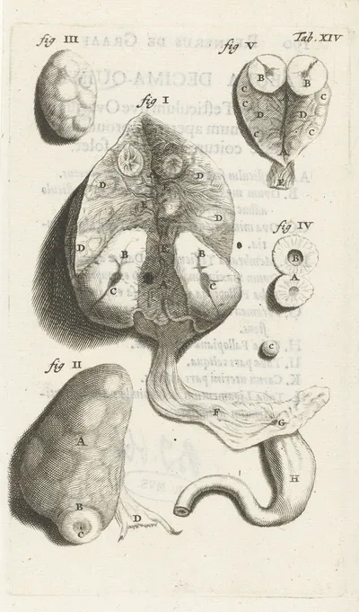
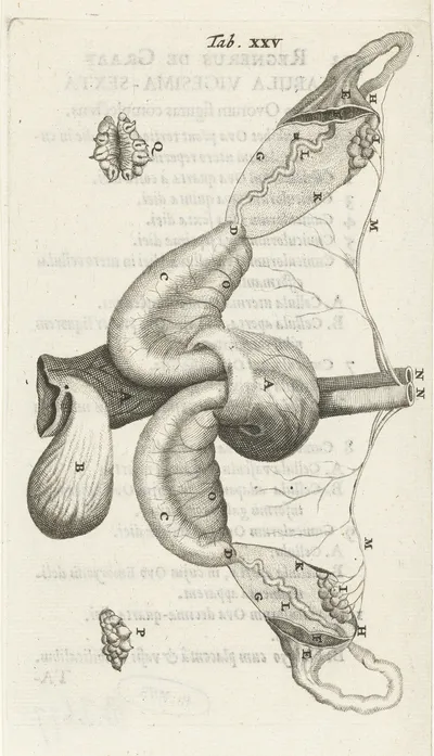
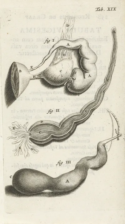
Anatomical illustration of the fallopian tube and ovaries (1672)
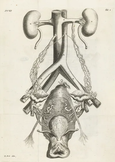
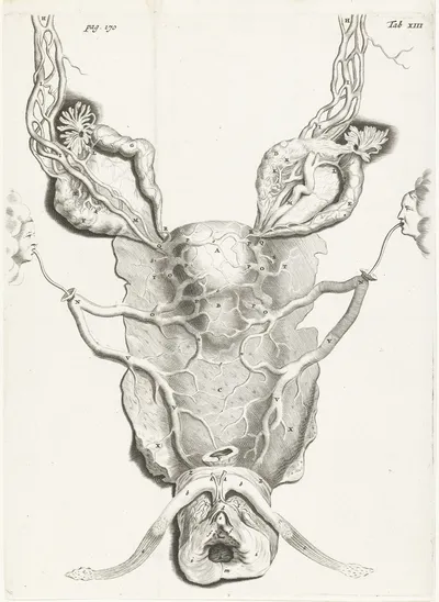
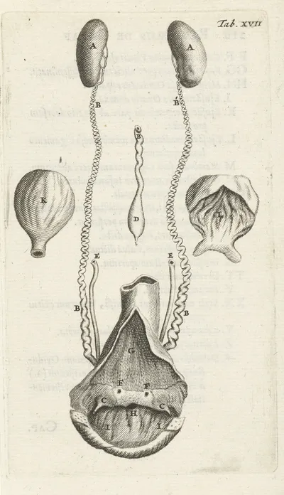
Anatomical illustration of the female reproductive organs and kidneys. (1672)
Anatomical illustration of the inside of the uterus (1672)
Anatomical illustration of the inside of the uterus, fallopian tube and ovaries (1672)
Anatomical illustration of the inside of the uterus. (1672)
Anatomical illustration of the ovaries and fallopian tubes (1672)
Anatomical illustration of the path of the egg in the uterus (1672)
Anatomical illustration of the vagina (1672)
Anatomical illustration of the vagina and the uterus (1672)
Anatomical illustration of the vagina and the uterus. (1672)
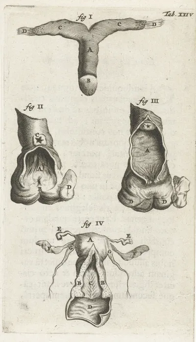
Anatomical illustration of various female genitalia. (1672)
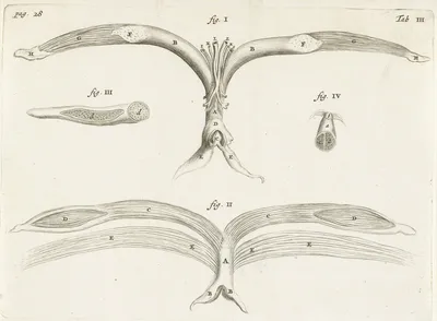
Anatomical image of a sphincter (1672)
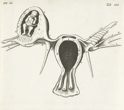
Anatomical image of an ectopic pregnancy (1672)
Anatomical image of an ectopic pregnancy. (1672)
Anatomical image of female genitalia. (1672)
Anatomical image of the uterus in enlarged form (1672)
Anatomical image of the uterus in enlarged form. (1672)
Anatomical representation of various female genitalia (1672)
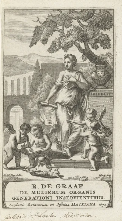
De mulierum organis generationi inservientibus tractatus novus – Title Page (1672)
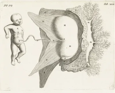
Fetus still connected to the uterus by the umbilical cord (1672)